

Doppler diagnostics
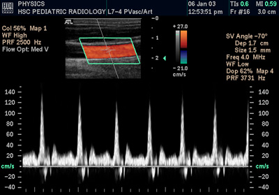 The phenomenon of echo Doppler ultrasound forms the basis of ultrasound diagnosis in vascular neurology. Its enables the unfolding of the vessel wall and quantitative assessment of flow velocity and its direction. Recent years have seen rapid development of these techniques. The introduction of color Doppler, power Doppler and Duplex and Triplex systems, significantly expands the diagnostic possibilities. With these new applications received non-invasive techniques to help assess vascular extra-and intracranial and monitor flow rates and collateral circulation.
Doppler effect - the phenomenon observed for the waves, involving the formation of the difference frequency wave sent by the source and recorded by the observer moves relative to the source wave. For waves that spread in the center, such as sound waves, the effect depends on the speed of the observer and the source terms of the medium in which these waves to propagate. In the case of waves propagating without the participation of a material, such as light in a vacuum (electromagnetic waves in general), the significance of the difference in speed is only the source and the observer.
Using the Doppler effect in medical diagnostics.
In the diagnostic imaging studies, valuable information is not only the shape of anatomical structures, but also the direction and speed of movement of tissue. Movement of body fluids such as blood can be monitored by measuring changes in frequency and phase of sound waves reflected from the flowing liquid. To improve conventional ultrasound apparatus was the introduction of Doppler ultrasound. If the ultrasound head can not only record the sound emitted echo delay, but also its height or phase, then the diagnostic image can illustrate the conventional color body movement.
Communicates
How to find us?
Click here to view the map
Dane firmowe
PREMIUM HOUSE KOŁŁATAJA 5Cneurochirurg.opole.plŁątka i Partnerzy
- Lekarze Neurochirurdzy sp.p.Kołłątaja 5C/13, 45-064 OpoleKRS 0000822292, NIP 7543264497
REGON 385244927-00011
BDO 0001/000460247mBank 81 1140 2004 0000 3302 7968 8364specjalisci.opole.plGlaubic Łątka s.c.Kołłątaja 5C/11, 45-064 OpoleNIP 7543156690, REGON 368230137mBank 88 1140 2004 0000 3402 7708 0732Specjalistyczna Prywatna Praktyka LekarskaŁątka DM s.c.Kołłątaja 5C/13, 45-064 OpoleNIP 7542841136, REGON 160054345mBank 41 1140 2017 0000 4802 0545 1697